Advanced dental diagnostics is defined as the clinical application of modern imaging and assessment technologies that detect oral disease and structural compromise earlier and more accurately than conventional examination alone. Technologies such as cone-beam computed tomography (CBCT), AI-assisted image interpretation, and Quantitative Percussion Diagnostics (QPD) now form the backbone of thorough oral health assessments in leading practices. These tools do not replace clinical judgement. They sharpen it, giving both patients and clinicians a clearer picture before treatment decisions are made. Understanding how these technologies work, and when each is most appropriate, is the foundation of informed dental care in 2026.
Advanced dental diagnostics explained: the core technologies
Modern dental diagnostics encompasses three distinct but complementary technology categories: three-dimensional volumetric imaging, artificial intelligence interpretation tools, and biomechanical integrity assessment. Each addresses a different diagnostic gap that conventional two-dimensional radiographs and visual examination leave open.
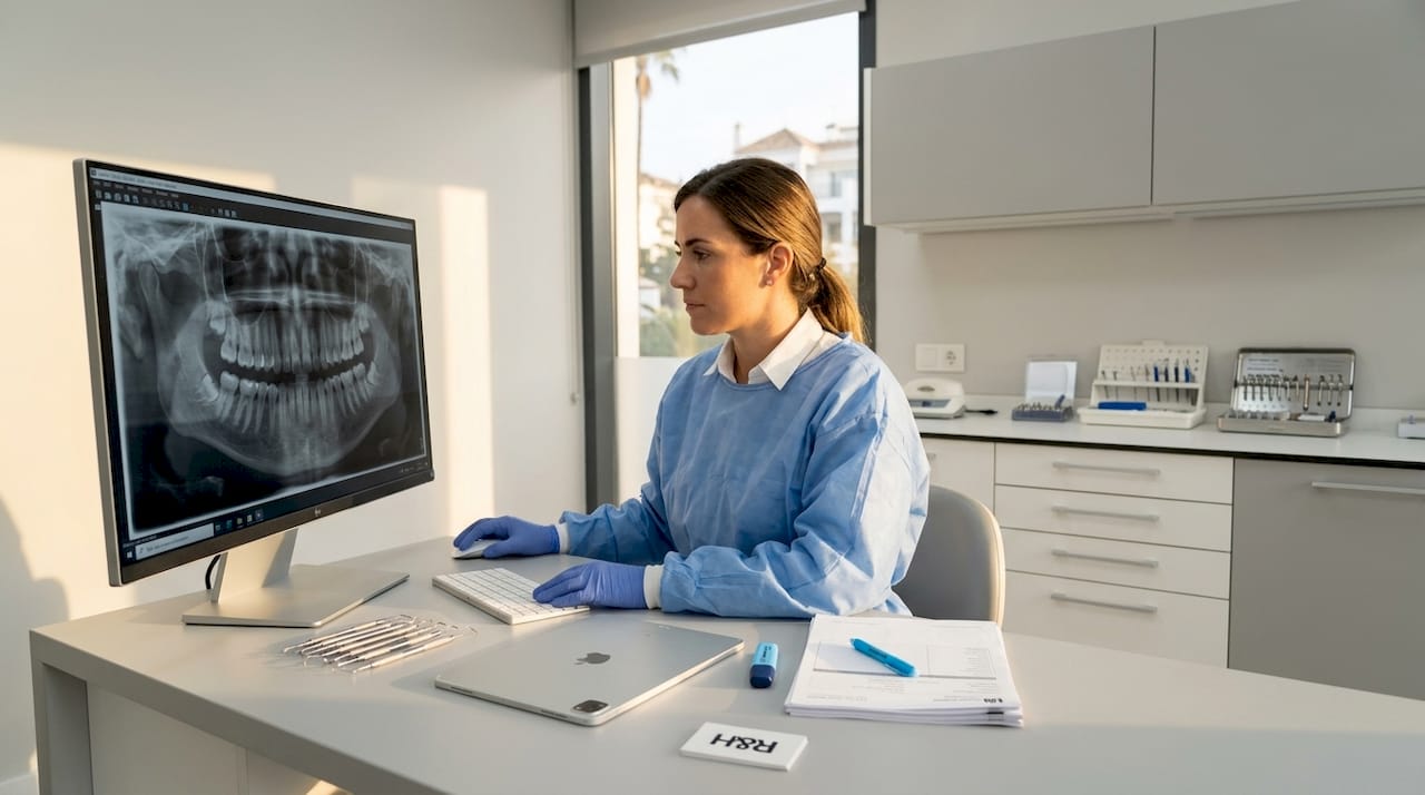
Dental X-rays detect damage and disease invisible during routine exams, yet even high-quality two-dimensional films miss early cracks, subtle bone loss patterns, and restoration failure at the adhesive interface. This limitation is precisely what advanced oral health assessments are designed to address.
The three named platforms most frequently cited in current clinical literature are Dentsply Sirona Smart View, the CephX AI platform, and the InnerView QPD system. Each targets a specific diagnostic challenge: Smart View addresses periapical pathology in CBCT scans, CephX automates cephalometric and 3D orthodontic analysis, and InnerView measures the biomechanical response of teeth and restorations. Together, they represent the current frontier of dental diagnostic technologies available in well-equipped clinics.
How does CBCT improve dental imaging accuracy?
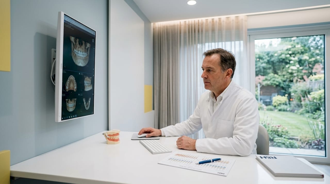
Cone-beam computed tomography produces a three-dimensional volumetric dataset from a single rotation of the X-ray source around the patient’s head. Unlike conventional two-dimensional periapical or panoramic films, CBCT renders individual voxels of tissue data, allowing the clinician to examine anatomy in axial, coronal, and sagittal planes simultaneously. This is particularly significant for implant planning, where bone width, height, and proximity to the inferior alveolar nerve must be measured with precision before any surgical intervention.
The clinical applications of CBCT extend well beyond implant planning. In endodontics, CBCT reveals missed canals, root fractures, and periapical lesions that flat films obscure. In orthodontics, it provides accurate skeletal measurements and airway assessment. In trauma cases, it identifies root fractures and alveolar bone injuries within minutes of the patient arriving at the chair.
Radiation dose is a legitimate consideration, and the American Dental Association endorses the ALARA principle for all dental imaging: exposure should be as low as reasonably achievable, with imaging intervals tailored to each patient’s clinical risk rather than fixed schedules. Modern CBCT units with small fields of view and digital flat-panel detectors deliver doses comparable to a full-mouth series of periapical films in many protocols.
AI is now integrated directly into CBCT review workflows. Dentsply Sirona’s Smart View Detect is the first FDA-cleared AI diagnostic aid for identifying periapical radiolucencies in CBCT scans, increasing detection sensitivity by approximately 46% without increasing false positives. This figure matters because periapical lesions missed on routine review are among the most common causes of delayed endodontic treatment and unnecessary tooth loss.
- Implant planning: CBCT measures bone volume and identifies anatomical hazards before surgery
- Endodontics: Reveals root fractures and periapical pathology invisible on two-dimensional films
- Orthodontics: Provides skeletal and airway data for accurate treatment planning
- Trauma assessment: Identifies alveolar and root injuries rapidly and non-invasively
Pro Tip: When asking your dentist about CBCT, request a small field-of-view scan limited to the area of clinical concern. This reduces radiation dose significantly compared to a full-arch volume and is sufficient for most single-tooth or localised assessments.
What role does artificial intelligence play in dental image interpretation?
Artificial intelligence in dental imaging operates through convolutional neural networks trained on large datasets of annotated radiographs. The network learns to recognise patterns associated with caries, bone loss, periapical pathology, and root fractures, then overlays its findings on the clinician’s screen as a decision-support layer. The clinician reviews, accepts, or overrides the AI suggestion. Responsibility for the final diagnosis remains entirely with the treating dentist.
A 2026 systematic review published in Cureus found that AI improves diagnostic efficiency and consistency for caries detection, bone loss assessment, and root-fracture interpretation. The same review noted significant methodological heterogeneity across studies and limited external validation, meaning results from one population or imaging system do not automatically transfer to another. This is not a reason to dismiss AI tools. It is a reason to use them within validated, cleared platforms rather than unregulated add-ons.
“AI in dental imaging is most valuable as a clinician-assisted tool that enhances efficiency and consistency. It does not yet have the autonomy to replace the experienced eye of a trained clinician, particularly in complex or ambiguous cases.”
CephX received FDA clearance for AI-powered 3D orthodontic imaging analysis, integrating with platforms including DEXIS and Greyfinch to automate cephalometric measurements and reduce inter-operator variability. For orthodontic patients, this means treatment plans built on consistent, reproducible data rather than measurements that vary between clinicians or appointments.
The practical implication for patients is straightforward. When a clinic uses cleared AI tools within its imaging workflow, you benefit from a second layer of pattern recognition that catches findings a tired or time-pressured clinician might overlook. What you should not expect is an AI system that diagnoses independently or communicates findings to you without clinical interpretation. The dentist remains the author of your diagnosis.
Quantitative percussion diagnostics: assessing structural integrity
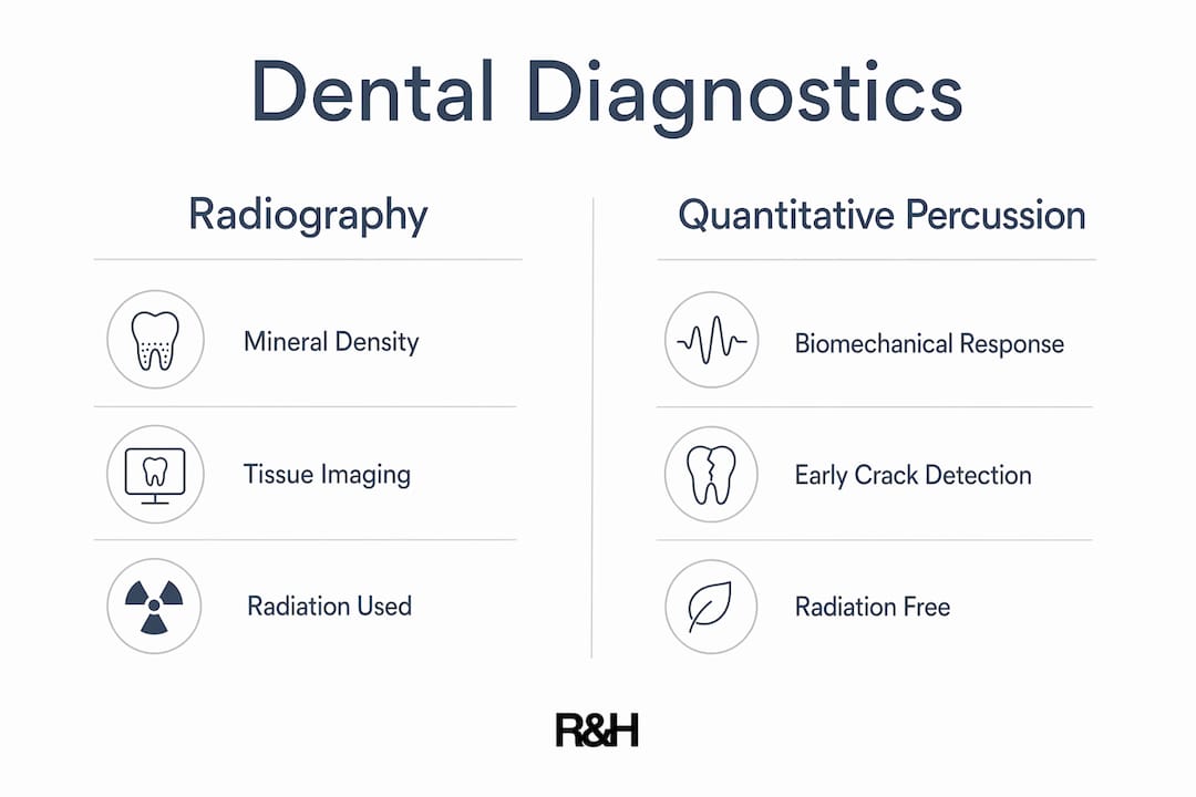
Quantitative Percussion Diagnostics (QPD), delivered through the InnerView system, represents a conceptually different approach to dental assessment. Rather than measuring mineral density or tissue contrast as radiography does, QPD measures the biomechanical response of the crown-tooth complex to a controlled percussive stimulus. The Energy Return Graph algorithm interprets micro-mobility, internal structural response, and adhesive interface behaviour to identify early cracks, adhesive degradation, and restoration instability.
The clinical value of this distinction is significant. Conventional radiographs may fail to detect early caries, cracks, and restoration failure until disease has advanced to a point where more invasive treatment becomes necessary. QPD identifies structural compromise at a stage when minimally invasive intervention is still possible, preserving more natural tooth structure and improving long-term prognosis.
The test itself takes under two minutes, produces no radiation, and requires no special patient preparation. This makes it suitable for routine integration into recall appointments, not just complex diagnostic workups.
| Feature | Radiography (2D/CBCT) | QPD / InnerView |
|---|---|---|
| What it measures | Mineral density and tissue contrast | Biomechanical response and micro-mobility |
| Radiation | Yes (low to moderate dose) | None |
| Early crack detection | Limited | Strong |
| Adhesive failure detection | Unreliable | Reliable |
| Test duration | 5-20 minutes | Under 2 minutes |
| Best for | Bone, periapical, anatomical assessment | Restoration integrity, fracture risk |
Pro Tip: If you have a crowned tooth that feels subtly different when biting but shows nothing on X-ray, ask whether QPD assessment is available. It is specifically designed to detect the kind of structural change that radiographs routinely miss.
Choosing the right diagnostic tool for the clinical scenario
No single technology answers every diagnostic question. The skill in modern dental diagnostics lies in selecting the right tool for the clinical scenario, or combining modalities where the question demands it.
For early interproximal caries detection, AI-assisted bitewing radiography with tools such as those integrated into DEXIS workflows offers improved sensitivity over unaided visual review. For implant planning, CBCT with a small field of view is the standard of care, and AI overlays like Smart View Detect add a further layer of periapical assessment. For a patient with a crowned molar showing no radiographic pathology but reporting vague biting discomfort, QPD provides information that no imaging modality can.
Combining modalities improves overall diagnostic confidence. A patient presenting for a full-arch implant assessment might receive CBCT for bone volume and anatomy, AI-assisted review for periapical status, and QPD for the remaining natural teeth to assess structural integrity before treatment planning begins. This layered approach reflects best practices in dental diagnostics as they are understood in 2026.
Patient-specific risk assessment governs imaging frequency. The ADA recommends customised imaging intervals based on individual oral health status rather than fixed annual schedules. A patient with active caries, periodontal disease, or complex restorations warrants more frequent imaging than a low-risk patient with a stable, well-maintained dentition.
Questions worth raising at your next consultation:
- Which imaging modalities are indicated for my specific clinical concern?
- Is the CBCT field of view limited to the area of interest to minimise dose?
- Are the AI tools in use FDA or CE cleared for the specific diagnostic task?
- Would QPD assessment add useful information for my crowned or heavily restored teeth?
How R&H Dental Marbella integrates advanced diagnostics into patient care
R&H Dental Marbella uses 3D CBCT imaging, AI-assisted analysis, and QPD assessment as part of a thorough diagnostic process that precedes any significant treatment recommendation. The clinic’s advanced dental technology includes an in-house digital laboratory and photography studio, which means diagnostic data flows directly into treatment planning and patient communication without being outsourced or delayed.
The clinical team brings 15 to 35 years of experience each, with dentists from Finland, New Zealand, Ireland, Portugal, and Spain. This breadth of training means that diagnostic findings are interpreted within a wide frame of clinical reference, not a narrow speciality silo. For patients who have relocated to Marbella or are visiting from abroad, this matters. You are not starting from scratch with an unfamiliar system. You are working with experienced clinicians who communicate clearly in English and explain findings in terms that support your decision-making.
The clinic’s approach to advanced oral health assessments reflects several clear commitments:
- Transparent pricing: Diagnostic costs are disclosed before any assessment begins, with no hidden fees
- Written guarantee: Treatment outcomes are backed by a formal written guarantee, not verbal reassurance
- Minimally invasive philosophy: Advanced diagnostics are used to catch problems early, reducing the need for extensive intervention
- Patient-led decisions: Findings are shared clearly, with time given for questions before any treatment is agreed
For patients considering dental implants in Marbella, the combination of CBCT planning and AI-assisted periapical review provides the most complete pre-surgical picture available in clinical practice today.
Key takeaways
Advanced dental diagnostics delivers its greatest value when imaging, AI interpretation, and biomechanical assessment are combined and matched to the specific clinical question at hand.
| Point | Details |
|---|---|
| CBCT transforms implant and endodontic planning | Three-dimensional volumetric data reveals anatomy and pathology invisible on two-dimensional films. |
| AI tools augment, not replace, clinical judgement | Cleared platforms like Smart View Detect and CephX improve detection consistency but require clinician oversight. |
| QPD detects what radiographs miss | Biomechanical assessment identifies early cracks and adhesive failure before radiographic signs appear. |
| Imaging intervals should be patient-specific | ADA guidance recommends customised schedules based on individual risk, not fixed annual routines. |
| Combining modalities improves diagnostic confidence | Layering CBCT, AI review, and QPD provides a more complete clinical picture than any single tool alone. |
Our perspective on diagnostics-led dentistry
What we have observed over years of clinical practice is that the most consequential improvements in patient outcomes do not come from new materials or techniques alone. They come from finding problems earlier. A crack identified by QPD before it propagates to the pulp is a tooth saved with an onlay rather than lost to extraction. A periapical lesion caught by Smart View Detect at two millimetres is a root canal rather than an implant. The difference in cost, recovery, and long-term prognosis is substantial.
We are also honest about the limits of these tools. AI platforms are only as reliable as the datasets they were trained on, and clinicians need to verify that any AI add-on is cleared for the specific diagnostic task it is being used for. CBCT is not appropriate for every clinical question, and dose justification matters. QPD is a complement to radiography, not a substitute for it.
What we encourage patients to do is ask questions. Ask which tools are being used, why they are indicated for your situation, and what the findings mean for your treatment options. An informed patient is a better partner in care, and better partners in care tend to achieve better outcomes. That is not a platitude. It is what the evidence consistently shows.
— R&H Dentists
Discover advanced diagnostics at R&H Dental Marbella
R&H Dental Marbella offers thorough diagnostic assessments using 3D CBCT imaging, AI-assisted analysis, and QPD structural integrity testing, all within a single appointment. Our experienced English-speaking team explains every finding clearly, so you leave understanding your oral health rather than simply holding a treatment plan.
Whether you are an expat living on the Costa del Sol or visiting Marbella for specialist care, our transparent pricing means you know the cost of your diagnostic assessment before you begin. There are no surprises, and no pressure. If you would like to discuss which diagnostic approach is right for your situation, we welcome you to book a consultation with our team at your convenience.
FAQ
What is CBCT and when is it used in dentistry?
Cone-beam computed tomography (CBCT) is a three-dimensional imaging system that produces volumetric data of teeth, bone, and surrounding anatomy. It is used for implant planning, endodontic assessment, orthodontic analysis, and trauma evaluation where two-dimensional radiographs are insufficient.
How does AI improve dental diagnostics?
AI tools trained on large radiographic datasets improve the consistency and sensitivity of findings such as caries, bone loss, and periapical lesions. A 2026 systematic review confirmed AI’s value as a decision-support tool, though clinician oversight remains necessary for all final diagnoses.
What is Quantitative Percussion Diagnostics (QPD)?
QPD is a radiation-free assessment that measures the biomechanical response of a tooth or restoration to a controlled percussive stimulus. The InnerView system uses Energy Return Graph algorithms to detect early cracks, adhesive failure, and micro-mobility that radiographs cannot reliably identify.
How often should dental X-rays be taken?
The ADA recommends patient-specific imaging intervals based on individual oral health risk rather than fixed annual schedules. Patients with active disease or complex restorations may require more frequent imaging than those with stable, low-risk dentitions.
Can advanced diagnostics reduce the need for invasive treatment?
Yes. Early detection through CBCT, AI-assisted review, and QPD allows clinicians to intervene at a stage when minimally invasive options such as onlays, targeted endodontics, or adhesive repair remain viable, preserving more natural tooth structure and improving long-term prognosis.
.png)